Introduction
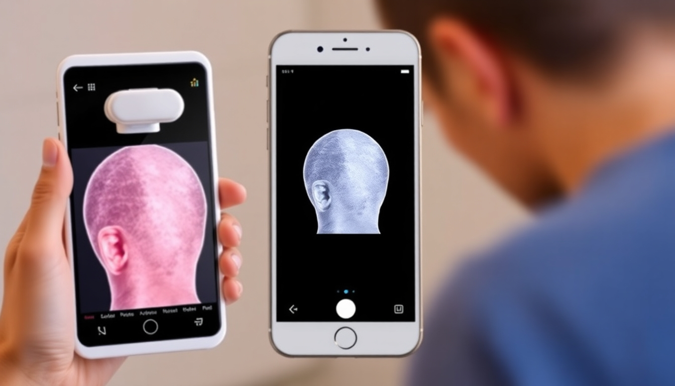
In 2025, the hair‑care market is crowded with peptide serums, prebiotic scalp treatments and an expanding array of at‑home scalp devices. Consumers and small clinics want reproducible, visual evidence that a product or device is creating meaningful change. "Pixels to Proof" is a practical, consumer‑friendly 6‑week split‑scalp protocol designed to photometrically validate changes in scalp condition while minimizing complexity.
What This Protocol Will Do — and What It Won't
- Do: Provide a repeatable, low‑cost approach to capture and compare visual changes between two scalp regions (treated vs. control) using consistent photography, simple scoring and accessible analysis.
- Do: Help you surface trends in redness, flaking, sheen, and perceived hair density.
- Don't: Substitute for clinical trials, diagnostic medical advice, or laboratory biomarker testing.
Why Use a Split‑Scalp Design?
A split‑scalp design (e.g., left vs. right, anterior vs. posterior) controls for between‑person variability because each participant acts as their own control. This design increases sensitivity to detect subtle, localized changes during a short timeframe like six weeks.
Science Snapshot: Peptides, Prebiotics & Devices
- Peptide serums: Short chains of amino acids designed to signal or support processes like keratin synthesis, barrier function or microcirculation. Visual effects often include improved scalp texture and sheen.
- Prebiotic scalp treatments: Formulations that aim to support a balanced scalp microbiome by supplying substrates that favor beneficial microbes. Visual targets include reduced flaking and less visible inflammation.
- At‑home scalp devices: Devices (LED, microcurrent, massage, microneedling) that can influence circulation, inflammation or transdermal delivery. Visual outcomes are typically supportive rather than dramatic.
Equipment & Setup — Consumer‑Friendly and Repeatable
To ensure photometric consistency prioritise reproducible lighting, camera placement and subject positioning. Here’s a practical equipment list with low‑cost options:
- Smartphone with a good camera (latest iPhone, Pixel, Samsung) or a compact digital camera.
- Sturdy tripod or phone mount to fix camera distance and angle.
- Neutral, non‑reflective background (plain wall or backdrop cloth).
- Two identical soft lights (daylight LED panels at ~5500K) or a single window with consistent time of day.
- Color reference card or gray card for white balance calibration.
- Marker (small tape on chair/floor) to keep head position consistent.
- Notebook or spreadsheet template for daily logs.
Illustrations to Use In Your Report
When publishing results include clear images with keyword‑rich alt attributes for SEO. Example image tags you can include in blog posts:
6‑Week Split‑Scalp Protocol — Detailed
This step‑by‑step timeline gives daily and weekly actions so results are comparable and defensible.
Pre‑Study Preparation (1 Week)
- Select volunteer(s) and obtain informed consent to photograph and publish images if results will be shared publicly.
- Decide your split approach: left vs. right, frontal vs. occipital, or superior vs. inferior. Mark sides A (treatment) and B (control).
- Standardize background and lighting. Place markers for where the person sits and where the camera is mounted.
- Measure a baseline: capture high‑resolution photos (top, 45°, and side views) and complete a baseline symptom/score sheet (redness, flaking, oiliness, perceived density — each 0–5).
- Ask participants to stop using any other targeted scalp treatments 48–72 hours before baseline if safe and practical. Keep shampoos/supplements stable during the trial.
Day 0 — Baseline Capture
- Capture images using the color/gray card in frame. Take at least three angles: vertex (top), 45° oblique and direct side.
- Record camera model, focal length, distance from scalp and lighting conditions.
- Assign product/device use instructions for side A only. Document application volume, frequency and device settings.
Weeks 1–2 — Initiation
- Apply product or device according to established instructions for side A. Keep side B on participant's normal maintenance routine, avoiding additional actives.
- Capture photos twice per week if possible (same days each week). Keep a daily log for symptoms and any reactions.
- At end of week 2, do a midpoint inspection for early irritation. If irritation occurs, stop treatment and consult a clinician.
Weeks 3–4 — Midpoint
- Continue the protocol. Capture standardized photos on the same days as weeks 1–2.
- At week 4, perform a structured mid‑trial scoring session (three blinded reviewers rate photos on redness, flaking, sheen, density using 0–5 scale).
- Adjust participant behaviors if necessary (e.g., reduce device intensity if mild sensitivity is observed).
Weeks 5–6 — Final Phase
- Complete the final two weeks of treatment per original schedule.
- At week 6 capture final high‑quality images using the baseline workflow (color card, same angles, same camera settings).
- Collect final subjective symptom scores and blinded reviewer ratings.
Post‑Protocol Analysis
Compile baseline, mid and final images and scores. Use both visual inspection and simple quantitative methods described below to reach conclusions.
Photography Technical Tips (Practical Camera Settings)
- Use manual or pro mode when possible. Lock exposure, focus and white balance.
- ISO: 100–200 to minimize noise.
- Aperture: f/4–f/8 for sufficient depth of field on compact cameras; smartphone apps handle depth automatically.
- Shutter speed: fast enough to avoid motion blur — 1/125s or faster if handheld, though tripod use is recommended.
- Use RAW capture if available; otherwise, high‑quality JPG is acceptable if white balance and exposure are consistent.
Lighting & Color Calibration
- Use two soft light sources at 45° angles to the subject to reduce harsh shadows and provide even illumination.
- Avoid mixed light sources (sunlight + indoor bulbs) which create white balance shifts.
- Include a gray card in at least the first and last images each session for post‑capture white balance correction.
Image Management & Preprocessing
Organize your files logically: ParticipantID_Side_Date_Angle (e.g., P01_A_2025-05-01_vertex.jpg). Preprocess images consistently:
- Apply identical white balance and exposure adjustments using batch features in Lightroom, Snapseed, or a similar tool.
- Crop to match the same framing region across sessions. Tools like Photoshop, GIMP or mobile apps can help align and crop consistently.
- Keep an untouched archive of originals for auditing.
Non‑Technical Analysis Methods (Accessible)
- Side‑by‑side comparisons: present baseline vs. final images to blinded reviewers for preference and perceived change.
- Simple scoring: use a 0–5 scale for redness, flaking, oiliness, and perceived density. Average scores across reviewers.
- Trend charts: plot average scores over time to visualize directionality and speed of change.
Optional Technical Image Analysis (For Enthusiasts)
For users who want more quantitative measures, open‑source tools like ImageJ/Fiji can extract color and area metrics.
- Redness index: convert to Lab color space and track changes in the a* channel (green–red axis). An increase in a* indicates more redness.
- Flake coverage: threshold on a brightness or color channel to detect white/gray flakes and compute percent area covered.
- Perceived density proxy: compute scalp contrast by measuring the ratio of dark pixels (hair) to background scalp pixels in a standardized crop.
- Automated alignment: use feature matching to align images and subtract baseline from final to highlight pixel‑level differences.
Scoring Rubric Example (0–5)
- Redness: 0 = none, 1 = trace, 2 = mild, 3 = moderate, 4 = marked, 5 = severe.
- Flaking: 0 = none, 1 = minimal, 2 = mild/localized, 3 = moderate, 4 = widespread, 5 = severe coverage.
- Sheen/Texture: 0 = dull, 5 = high healthy sheen (use same lighting to judge).
- Perceived density: 0 = very sparse, 5 = very dense (assessed with consistent hair parting).
Interpreting Results — Practical Guidance
- Look for directional consistency across modalities: if blinded reviewers, subjective scores and pixel metrics all trend the same way, confidence increases.
- Small absolute changes can be meaningful if consistent and reproducible across participants or repeated trials.
- Document adverse events. Visual improvement is not worth ongoing irritation or allergic reactions.
Common Pitfalls & Troubleshooting
- Inconsistent lighting: leads to false positives/negatives. Recalibrate and retake images if necessary.
- Different hair styling or product residue: require participants to wash hair prior to imaging or maintain a consistent styling routine.
- Camera auto adjustments: disable auto HDR, auto exposure or auto white balance.
- Participant non‑adherence: keep a simple daily log and periodic check‑ins to ensure protocol fidelity.
Ethics, Safety & Privacy
- Obtain explicit consent to photograph and publish images. Offer the option to anonymize images (crop face out or blur identifiable features).
- Advise participants to stop treatment and seek medical advice if they develop significant irritation.
- Store images and logs securely, respecting privacy regulations in your jurisdiction.
Publishing Findings — SEO & Shareability Tips
- Use descriptive, keyword‑rich headlines and subheadlines (e.g., "6‑Week Split‑Scalp Trial: Peptide Serums vs. Control").
- Include alt text on all images with target keywords like "peptide serums", "prebiotic scalp treatments" and "at‑home scalp devices" for discoverability.
- Provide clear captions and a gallery of baseline vs. final images with a short methodology description for transparency.
- When linking to product pages, use natural anchor text that matches user intent (e.g., "try Eelhoe peptide serums" rather than generic "click here").
Case Example (Hypothetical)
Participant P01 used a peptide serum on side A and standard shampoo on side B. Baseline redness was 3/5 bilaterally. At week 4 redness on side A fell to 1.5/5 and flaking decreased from 3/5 to 1/5. Blinded reviewers preferred side A in 4 of 5 final comparisons. Pixel analysis of the a* channel showed an average 12% reduction in mean a* on side A versus 2% on side B. These convergent data increase confidence that the peptide serum contributed to reduced visible inflammation and flaking for this participant.
Appendix: Printable Checklist
- Pre‑study: consent form, set up camera/tripod, gray card, background.
- Day 0: baseline photos (vertex, 45°, side), symptom scores, assign sides.
- Weekly: capture photos same days, log product use, note any reactions.
- Week 4: midpoint blinded scoring.
- Week 6: final photos, final scores, compile blinded reviewer results and pixel metrics if used.
- Post: store raw images, publish results with transparent methods and alt‑tagged images.
Frequently Asked Questions
- How many participants do I need? For consumer self‑experiments, one participant with good documentation can be informative. For broader claims, recruit multiple participants to increase reliability.
- Can lighting truly be standardized at home? Yes — pick a time and place with controllable LED lights or use a pair of daylight LED panels and keep them in the same positions for every session.
- Will smartphone photos be good enough? Absolutely — modern smartphones capture high‑quality images. The key is consistency in camera positioning and settings.
Further Reading & Tools
- ImageJ/Fiji (open source): for image segmentation and color analysis.
- Lightroom or Snapseed: batch white balance and exposure correction.
- Clinical photography guidelines (general search for dermatologist or cosmetic imaging best practices).
Conclusion — From Pixels to Practical Decisions
Translating visual impressions into reproducible evidence is within reach for motivated consumers and small clinics. A rigorous 6‑week split‑scalp protocol emphasizes consistency in photography, structured scoring, and straightforward analysis. Whether your goal is to decide whether to keep using a peptide serum, evaluate a prebiotic scalp treatment, or validate an at‑home scalp device, this approach turns subjective impressions into actionable visual data.
Try Products That Pair Well With This Protocol
If you want to test formulations engineered for visible scalp improvements, check out Eelhoe's range of targeted solutions. Explore their peptide serums, prebiotic scalp treatments and compatible at‑home scalp devices and use this 6‑week split‑scalp protocol to capture your own pixels to proof. If you’re ready to test and want curated products to match this methodology, visit Eelhoe to browse formulations developed for measurable scalp care outcomes.
Final Checklist Before You Start
- Informed consent and privacy plan for images.
- Set up consistent camera and lighting positions and mark them.
- Prepare product dosing and device settings for side A only.
- Create a logging spreadsheet for daily notes and weekly image uploads.
- Schedule blinded reviewers for midpoint and final comparisons if possible.
Good luck with your trial. With thoughtful preparation, this split‑scalp photometric protocol will help you move from anecdote to evidence — from pixels to proof.


Leave a comment
All comments are moderated before being published.
This site is protected by hCaptcha and the hCaptcha Privacy Policy and Terms of Service apply.